Pterygium Surgery
Pterygium surgery removes abnormal tissue growth from the surface of the eye and replaces it with healthy tissue to reduce the chance of recurrence. This procedure is recommended when a pterygium causes persistent symptoms or threatens vision.
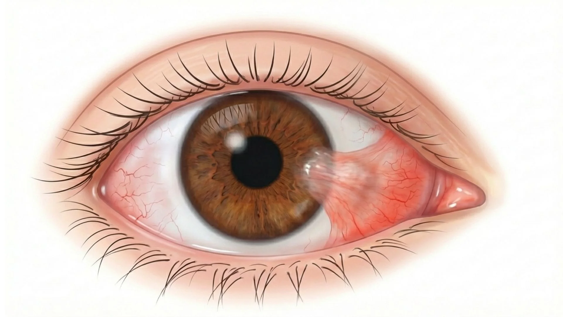
Understanding Pterygium
A pterygium is a wing-shaped growth of fleshy tissue that extends from the conjunctiva (the clear membrane covering the white of the eye) onto the cornea. While not cancerous, a pterygium can cause significant discomfort and visual problems if left untreated.
Pterygia are strongly associated with sun exposure and are common in tropical and subtropical climates, including Singapore. They are sometimes called "surfer's eye" due to their association with outdoor activities.
Problems caused by pterygium include:
Chronic redness and irritation
Foreign body sensation
Dryness due to uneven tear distribution
Astigmatism as the growth distorts the cornea
Visual obstruction if it grows across the pupil
When Surgery Is Recommended
Surgery may be advised when:
The pterygium causes persistent discomfort despite lubricating drops
Growth is progressing toward the central cornea
Vision is affected by induced astigmatism
The pterygium is cosmetically bothersome
It interferes with contact lens wear
The American Academy of Ophthalmology provides information about pterygium and its management.
The Surgical Procedure
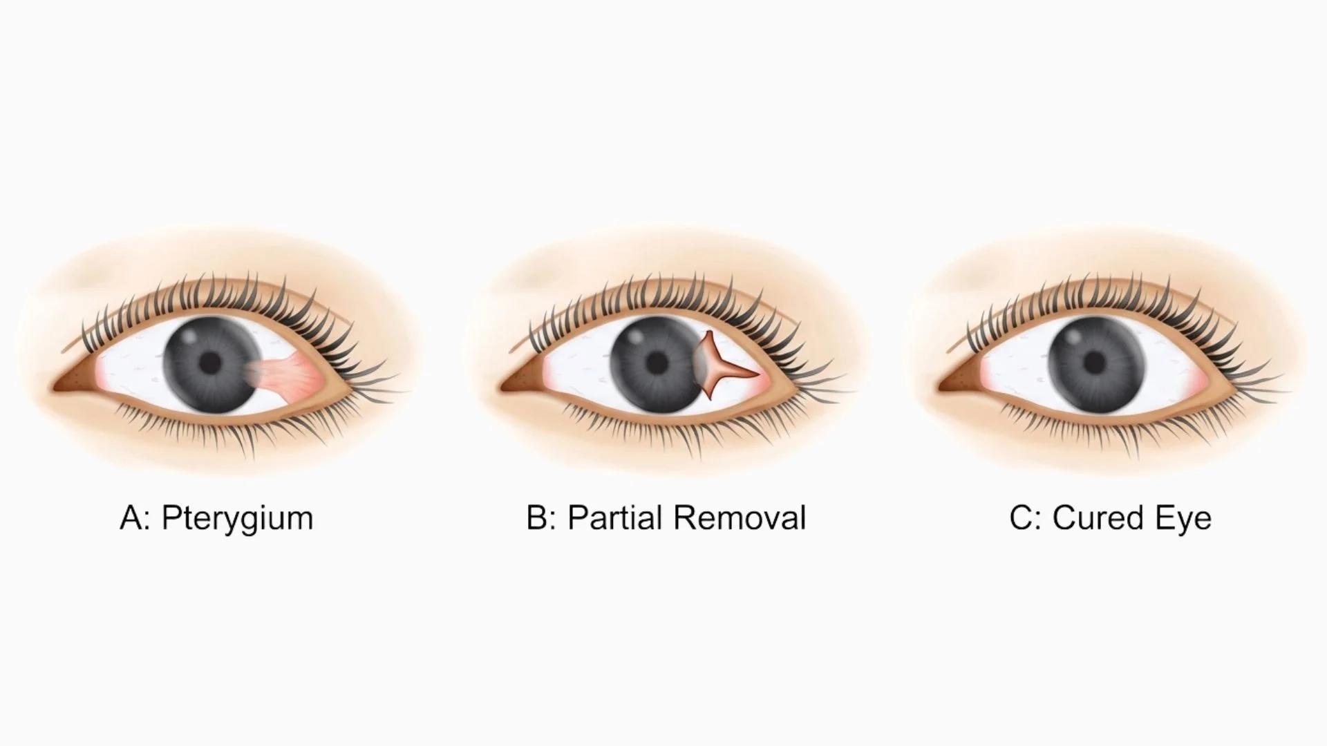
Modern pterygium surgery involves removing the abnormal tissue and transplanting healthy tissue to cover the area:
Pterygium Excision with Conjunctival Autograft:
The pterygium is carefully dissected from the cornea and sclera
A thin piece of healthy conjunctiva is taken from under the upper eyelid
This graft is secured over the bare area using tissue glue or fine sutures
The procedure takes approximately 30-45 minutes
Performed under local anaesthesia as a day procedure
This technique has significantly reduced recurrence rates compared to older methods that simply removed the pterygium without grafting.
What Results Can Be Expected
With conjunctival autograft technique:
Recurrence rates are typically 5-10% compared to up to 50% with simple excision
Most patients experience significant symptom relief
Redness and irritation usually improve substantially
Vision often improves as corneal distortion resolves
The eye typically appears much whiter once healed
Full healing takes several weeks, and the final appearance improves over 2-3 months.
Potential Risks
Recurrence remains possible despite grafting, particularly in younger patients
Graft displacement in the early healing period
Scarring of the cornea where the pterygium was attached
Infection is uncommon but requires prompt treatment
Persistent redness during the healing phase
Granuloma formation at the surgical site
Diplopia (double vision) is rare and usually temporary
Long-term prevention: Wearing UV-protective sunglasses and a wide-brimmed hat outdoors is essential to reduce the risk of recurrence and protect your other eye.
Medical Disclaimer: This information provides general guidance about pterygium surgery and should not replace professional medical advice. Individual outcomes depend on the size and extent of the pterygium, as well as healing response. Recurrence is possible despite modern surgical techniques. Please consult with our ophthalmologists for assessment and recommendations specific to your situation.
Pre-Operative Care
Discuss all medications with your surgeon
Arrange transportation home as your eye will be patched
No special fasting is required for local anaesthesia
Avoid wearing contact lenses for several days before surgery
The procedure is performed as a day case
Post-Operative Care
Seek attention if you experience: Severe pain, significant vision decrease, increasing discharge, or if you notice the graft appears displaced.
