Key Takeaways
Glaucoma is a group of eye diseases that slowly damage the optic nerve—the “cable” carrying information from the eye to the brain.
Most people experience no symptoms until significant vision loss occurs, and vision loss from glaucoma is usually irreversible, which is why glaucoma is often called the "silent thief of sight"
Sudden eye pain, a red eye, blurred vision or halos around lights—especially with headache, nausea or vomiting—needs urgent medical attention as it could be a sign of acute glaucoma where the eye pressure rises significantly over a short time.
In a 2025 study of Singaporeans aged 60 years and older, 5.1% (or 1 in 20 people) had glaucoma. (Gupta et al, 2025)
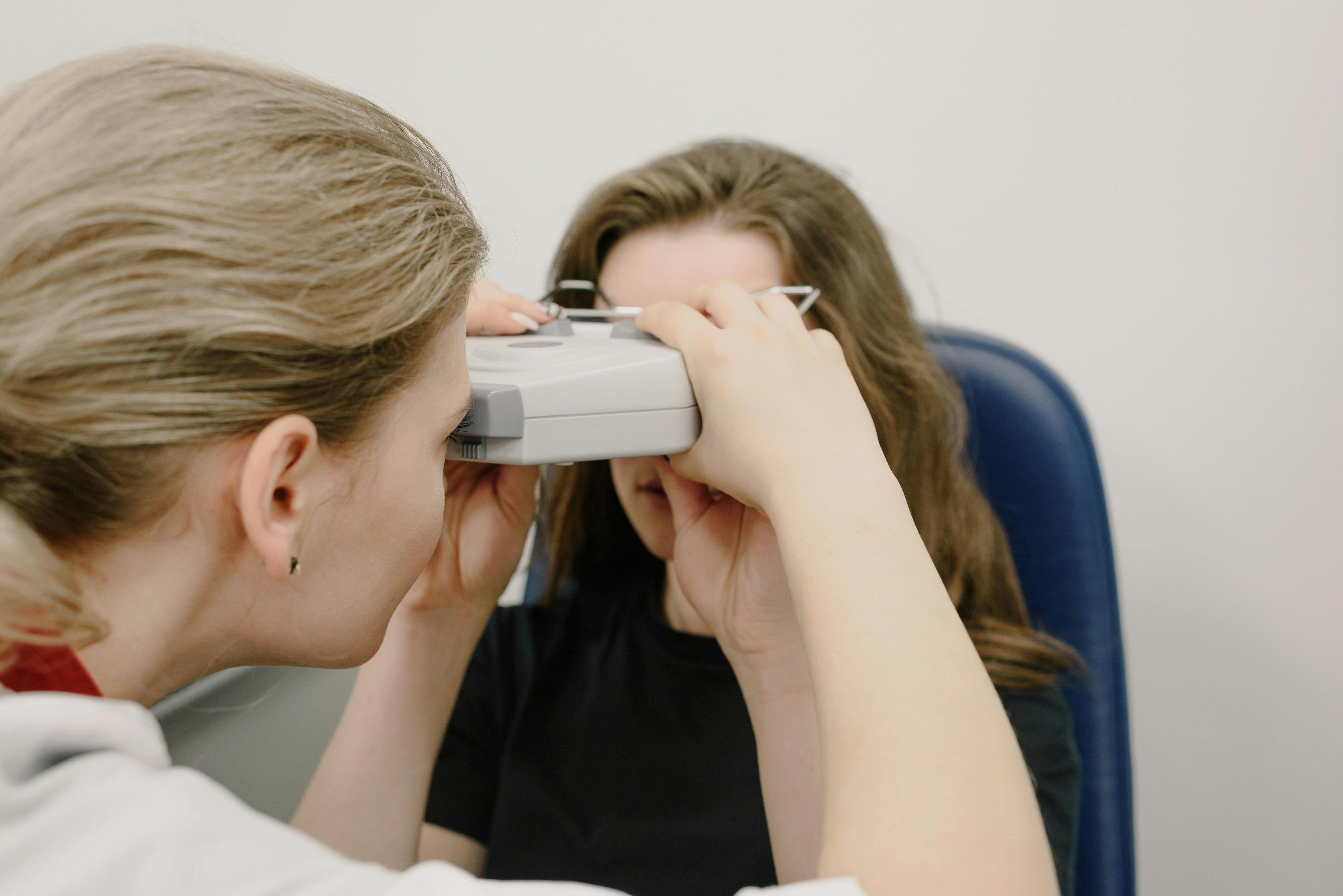
A proper glaucoma assessment requires several tests, and may include eye-pressure measurement (tonometry), examination of the structures of the eye and the optic nerve, gonioscopy (looking at the eye’s drainage angle), visual-field testing (HVF), OCT (a cross-sectional scan) of the retinal nerve fibre layer (RNFL) and ganglion cell inner plexiform layer (GCIPL), and corneal thickness (pachymetry) testing.
Goal of glaucoma treatment: To lower eye pressure to a safe target level, slow or prevent further optic nerve damage, and preserve your remaining vision, as vision already lost to glaucoma cannot be restored. This can be done by using glaucoma medications, lasers or surgery.
For selected people with open-angle glaucoma or high eye pressure, selective laser trabeculoplasty (SLT) is an effective, generally well-tolerated treatment that can lower eye pressure and reduce dependence on daily eyedrops.
For people with close-angle glaucoma, laser peripheral iridotomy or cataract extraction may be needed to help widen the drainage angle and improve the flow of fluid out of the eye.
Trans-scleral cyclophotocoagulation (TCP) uses laser energy delivered through the white of the eye to reduce fluid production and lower eye pressure, particularly in selected cases of difficult-to-control glaucoma.
Glaucoma surgery may be an option for selected patients whose eye pressure is not adequately controlled with eyedrops, who cannot tolerate glaucoma medications, or who wish to reduce their reliance on eyedrops. The most appropriate treatment depends on the severity of glaucoma, target eye pressure, and individual patient needs.
What is glaucoma?
Glaucoma is a group of diseases that damage the optic nerve. The optic nerve connects the eye to the brain, so damage can cause permanent loss of vision. This makes early detection and intervention critical. Catching glaucoma early, when we can still preserve vision, makes an enormous difference to long-term eye health.
Glaucoma is often associated with high eye pressure, also called intraocular pressure (IOP).
What is intraocular pressure (IOP)?
Think of your eye like a plumbing system. Fluid, called aqueous humour, which nourishes the eye tissues and contributes to the IOP within the eye, is continuously produced by the ciliary body within the eye, and needs to drain out through a drainage channel called the trabecular meshwork. When the drainage system isn't working efficiently, pressure builds up inside the eye. Over time, this elevated pressure damages the delicate nerve fibres of the optic nerve, which affects your peripheral vision at the initial stages and eventually progresses to affect your central vision if left untreated.
How Common is Glaucoma in Singapore
Singapore’s population is ageing and includes several ethnic groups with different patterns of glaucoma risk. Recent local population-based from the PIONEER study, which examined adults aged 60 to 100 years between 2017 and 2022 found that 5.1% of participants had glaucoma. This included:
3.4% with primary open-angle glaucoma (POAG).
0.7% with primary angle-closure glaucoma (PACG).
1.0% with secondary glaucoma, meaning glaucoma linked to another eye or medical problem.
The study estimated that about 57,800 Singaporeans aged 60 years and older were living with glaucoma, with a projected increase of 43% to about 85,800 by 2040 (Gupta et al, 2025).
Older global studies also show that glaucoma becomes more common with age (Tham et al, 2014).
The main types of glaucoma
Primary vs Secondary Glaucoma
Primary glaucoma refers to when the disease happens on its own, usually due to genetic traits, aging or the natural physical shape of the eye. There is no other underlying disease or external cause, and it is the most common form of glaucoma.
Secondary glaucoma refers to when the high eye pressure and nerve damage are a direct effect of an underlying issue, such as eye inflammation, an injury, previous eye surgery, steroid treatment, or changes such as pseudoexfoliation or pigment dispersion. To manage secondary glaucoma, the underlying cause also has to be managed.
Open-Angle vs Angle-Closure
Glaucoma can also be classified into open-angle or close-angle glaucoma. The front part of the eye has a drainage angle where fluid leaves the eye. In angle-closure disease, this angle is narrow or closed. This can be contributed by anatomical factors (e.g a shorter eye length or a thick lens), genetic factors, or other secondary factors.
To determine whether a patient has open-angle or angle-closure disease, the doctor may do tests including a gonioscopy or an anterior segment optical coherence tomography.
Distinguishing open-angle from angle-closure glaucoma is important because they may present differently and require different treatment approaches.
In Primary Open Angle Glaucoma (POAG), the eye’s drainage angle looks open, but fluid does not leave the eye as easily as it should because the drainage tissue isn't functioning properly. This often leads to a gradual, usually painless and symptomless increase in eye pressure, which is why timely detection, appropriate pressure control and ongoing monitoring can help preserve vision.
Unlike POAG, angle-closure glaucoma can develop suddenly and become a medical emergency. When eye pressure rises rapidly, patients may experience severe eye pain, blurred vision, headache, nausea, and vomiting. An acute attack can cause permanent vision loss within hours if not treated promptly, so immediate medical attention is essential.
Angle-closure disease exists on a spectrum, ranging from a narrow drainage angle without damage to established glaucoma with permanent optic nerve damage.
Primary Angle Closure Suspect (PACS): The drainage angle is narrow, putting the patient at risk of blockage. However, eye pressure is normal, there is no scar tissue (peripheral anterior synechiae, or PAS), and there is no optic nerve damage.
Primary Angle Closure (PAC): The narrow angle has begun to cause problems. Eye pressure may be elevated, and/or scar tissue (PAS) may have formed where the iris sticks to the drainage area. However, there is no evidence of glaucomatous optic nerve damage yet.
Primary Angle-Closure Glaucoma (PACG): The narrow angle has caused damage to the optic nerve, often associated with raised eye pressure and/or scar tissue.
Angle-closure glaucoma is particularly relevant in Singapore and across Asia. The prevalence of primary angle closure glaucoma (PACG) is higher in Asians than Europeans and Africans, with over 80% of PACG worldwide in Asia (Tham et al, 2014; Cheng et al, 2014).
If there are signs of angle closure, laser peripheral iridotomy or cataract surgery may be recommended to help open the drainage angle and improve the flow of fluid out of the eye. If eye pressure is significantly elevated, medications may also be needed promptly to lower the pressure and reduce the risk of damage to the optic nerve. The most appropriate treatment depends on the severity of angle closure and the individual patient's eye condition.
Normal-tension glaucoma
Normal-tension glaucoma refers to a condition where optic nerve damage occurs despite eye pressure being in what we'd typically consider the "normal" range. The diagnosis is based on the pattern of damage to the optic nerve and visual field, repeated pressure measurements and the exclusion of other causes.
Interestingly, up to 70% of open angle glaucoma in Asia occurs with normal or low intraocular pressure, compared to Western populations which reported a prevalence of around 30% amongst those with open angle glaucoma (Chen et al, 2020).
Treatment often still involves lowering eye pressure, but the target and any additional investigations must be tailored to the individual. Factors beyond simple pressure, such as reduced blood flow to the optic nerve, may be contributing, and may need to be managed.
Adult onset vs congenital and childhood/juvenile glaucoma
Glaucoma can occur at any age. However, adult-onset glaucoma is much more common than congenital or childhood/juvenile glaucoma.
Congenital glaucoma refers to a type of glaucoma that is present at birth (or developing within the first few months of life) due to an abnormal development of the eye. As an infant’s eye tissue is highly flexible, high pressure can cause the eye to stretch and enlarge (buphthalmos). Other common signs and symptoms of congenital glaucoma include excessive tearing, sensitivity to light, frequent blinking or squeezing of the eyelids, and cloudiness of the cornea. Childhood or juvenile glaucoma refers to glaucoma which develops later in childhood or adolescence (usually after age 3). There may be underlying developmental issues, genetics or secondary causes.
Symptoms: why glaucoma is often missed
Early glaucoma usually causes no symptoms. Reading and central vision may remain clear while peripheral vision is gradually affected. If one eye sees better than the other, it may compensate for the affected eye, making the loss of vision less noticeable. Because these changes occur slowly, the brain may also adapt, and patients may not realise that their vision is deteriorating until significant damage has occurred.
Symptoms in later stages of glaucoma may include:
Missing objects to the side: Gradual loss of peripheral (side) vision can make it harder to notice objects or people outside the direct line of sight.
Difficulty seeing in dim light: Some people may find it harder to navigate in poorly lit environments.
Difficulty reading or recognising faces: These problems may occur in advanced glaucoma when central vision becomes affected.
While the absence of symptoms does not rule out glaucoma, eye discomfort or blurred vision can also result from many other eye conditions and does not necessarily indicate glaucoma. A comprehensive eye examination is essential to determine the cause of symptoms and detect glaucoma early.
When to seek help immediately
A sudden rise in eye pressure can occur in acute angle-closure glaucoma, when the drainage angle becomes blocked abruptly. This can cause warning symptoms such as:
Eye pain.
Eye redness.
Blurred vision or coloured halos around lights.
Headache.
Nausea or vomiting.
Not all symptoms may be present, and an acute attack can sometimes be mistaken for a migraine, stomach illness, or another medical condition. If these symptoms occur, seek immediate medical attention at an eye clinic or emergency department. Do not wait for a routine appointment, as a sudden, severe rise in eye pressure can cause permanent vision loss within hours if treatment is delayed (Chan et al., 2025).
Who is more likely to develop glaucoma?
Several factors increase your risk of developing glaucoma, and understanding them can help you take proactive steps towards earlier or more frequent checks.
Risk factors include:
Older age.
A first degree relative (parent, brother, sister or child) with glaucoma.
Previously raised eye pressure, also called ocular hypertension.
A narrow or “occludable” drainage angle.
High myopia (short-sightedness).
Certain family and ethnic backgrounds. People of African descent have higher rates of POAG, while those of Asian descent have higher rates of angle-closure glaucoma (Cheng et al, 2014). In Singapore, the PIONEER study found that Malays and Indians had a higher prevalence of glaucoma compared Chinese (Gupta et al, 2025).
Previous eye injury, inflammation or surgery.
Long-term or repeated steroid use.
Conditions such as pseudoexfoliation or pigment dispersion.
Medical conditions that affect general vascular or metabolic health, e.g. diabetes, hypertension, hyperlipidaemia, smoking or obstructive sleep apnoea.
Screening in Singapore: who should be checked?
Screening means checking for the disease before symptoms develop. This is different from monitoring, which involves regular tests after glaucoma has been diagnosed or suspected.
There is no single screening schedule that is suitable for everyone. The frequency of eye examinations depends on your age, risk factors and eye health.
HealthHub and the Singapore National Eye Centre information encourages a baseline comprehensive eye examination at around age 40 and more frequent review when risk factors are present.
Your clinician will recommend an appropriate examination schedule based on your individual risk.
A practical guide is:
Routine screening: Arrange a baseline comprehensive eye examination and follow your clinician’s advice on subsequent checks, even if you have no symptoms.
Higher risk: You may need earlier or more frequent examinations if you have a strong family history of glaucoma, high eye pressure, high myopia, diabetes, narrow drainage angles, steroid exposure, or a history of eye injury or surgery.
If glaucoma is suspected or diagnosed: Follow your ophthalmologist’s recommended schedule for further tests and monitoring. This is disease monitoring rather than routine screening.
How is glaucoma assessed?
There is no single test that can diagnose glaucoma on its own. Diagnosis requires a combination of tests and the ophthalmologist combines several pieces of information to come up with a diagnosis.
At International Eye Cataract and Retina Centre (IECRC), we offer these tests as part of a comprehensive glaucoma assessment. If glaucoma is suspected, follow-up visits and additional tests may be arranged to confirm the diagnosis, assess disease severity, and plan appropriate treatment.
Eye-pressure measurement
Tonometry measures the pressure inside the eye, known as intraocular pressure (IOP).
This is done using either a puff of air (non-contact tonometry) or a gentle probe that touches the eye after numbing drops are applied (applanation tonometry).
A single IOP reading provides only a snapshot, as pressure can fluctuate throughout the day and between visits. Sometimes, a phasing test may be needed to measure IOP at different times of the day. This helps your doctor assess pressure patterns and identify fluctuations that may be missed during a single clinic visit.
IOP is the most important modifiable risk factor for glaucoma. Lowering IOP is the main way to slow disease progression and help preserve vision.
IOP is important, but it is only part of the picture. Normal IOP ranges from 6-21 mmHg. However, some people develop glaucoma even when their IOP is within the “normal” range. Other people have IOP but no signs of nerve damage. This is why other tests are required as part of a comprehensive evaluation for glaucoma.
Optic-nerve examination
The ophthalmologist examines the optic nerve using a slit-lamp microscope, which magnifies the structures inside the eye. A special lens is used to focus the light onto the optic nerve. The doctor will look for features suspicious of glaucoma, including thinning or notching of the nerve’s rim, asymmetry between the eyes, bleeding on the optic disc (also called a disc haemorrhage) and changes in the nerve-fibre layer.
The cup-to-disc ratio (CDR) describes the size of the central dip (the “cup”) relative to the entire optic nerve head (the “disc”). A large cup-to-disc ratio may suggest glaucoma, but some people naturally have larger optic nerve cups. Further tests are needed to determine whether glaucoma is present.
Gonioscopy
Gonioscopy is an examination where a special mirrored lens called a gonioscope is placed on the eye after placing numbing eyedrops on the eye, allowing the ophthalmologist to physically see all 360 degrees of the drainage system. It is considered the gold standard for determining whether the drainage angle is open or closed.
Anterior Segment OCT (ASOCT)
An Anterior Segment OCT (ASOCT) scan is a cross-sectional scan that can take an image of the drainage angle. It provides documentation and objective structural measurements of the drainage angle, but it cannot replace gonioscopy which is generally considered the gold standard.
Visual-field testing
A visual field test measures the area you can see while looking straight ahead. You will look at a central target while small lights flash at different locations around it, pressing a button whenever you see a light.
The test produces a map of your vision, helping detect areas of vision loss caused by glaucoma. It also helps assess the severity of vision loss and monitor for progression over time. Factors such as tiredness, difficulty understanding the test, cataracts, or unreliable responses can affect the results. Therefore, an abnormal or suspicious result may need to be repeated for consistency.
OCT scanning
Optical coherence tomography (OCT) is a non-invasive scan that produces detailed cross-sectional images of the optic nerve and retinal nerve layers, including the retinal nerve fibre layer (RNFL) and ganglion cell–inner plexiform layer (GCIPL). It measures their thickness to help detect early damage and monitor changes over time.
Corneal thickness (pachymetry)
This is a painless test that measures the thickness of the clear front surface of your eye (the cornea). Corneal thickness can affect eye pressure readings and pachymetry testing helps your ophthalmologist interpret the results more accurately when assessing your risk of glaucoma.
How is glaucoma treated?
The aim of glaucoma treatment is to lower eye pressure enough to reduce the risk of further optic nerve damage and preserve your remaining vision. Treatment cannot restore nerve fibres that have already been lost, but it can help slow or prevent further vision loss.
The target eye pressure varies for each person and depends on the extent of existing damage, how quickly the glaucoma is progressing, and overall health. Your ophthalmologist will determine the appropriate target and adjust treatment as needed. At International Eye Cataract and Retina Centre (IECRC), we offer all the following glaucoma treatments:
Eye-pressure lowering eyedrops
Different drops lower eye pressure in different ways and are usually classified according to how they work:
Prostaglandin Analogs (e.g., Bimatoprost, Tafluprost, Latanoprost):
How they work: Help fluid drain out of the eye.
Frequency: Usually once a day (often at night).
Common side effects: Eye redness, longer/darker eyelashes, and gradual darkening of the iris or eyelid skin, droopy lids, and sunken eyes.
Beta-Blockers (e.g., Timolol):
How they work: Reduce the amount of fluid your eye produces.
Frequency: Usually twice a day.
Common side effects: Fatigue, reduced heart rate or blood pressure, shortness of breath in people who have a history of asthma or other respiratory diseases; use with caution if you have asthma or heart conditions.
Alpha-Adrenergic Agonists (e.g., Brimonidine):
How they work: Reduce fluid production and increase drainage.
Frequency: Two to three times a day.
Common side effects: Dry mouth, fatigue, or eye irritation/allergic reactions.
Carbonic Anhydrase Inhibitors (e.g., Dorzolamide, Brinzolamide):
How they work: Decrease fluid production in the eye.
Frequency: Two to three times a day.
Common side effects: Temporary blurred vision, eye discomfort or a bitter taste in the mouth.
Rho Kinase (ROCK) Inhibitors (e.g., Netarsudil):
How they work: Improve fluid outflow.
Frequency: Once daily.
Common side effects: Eye redness or dryness.
Combination Drops (e.g., Dorzolamide/Timolol, Brimonidine/Timolol):
How they work: Combine two different mechanisms in a single bottle if you need extra pressure control.
Preservative-Free (PF) Options (Available for several classes above, e.g., Preservative-Free Tafluprost, Bimatoprost, or Dorzolamide/Timolol):
How they work: They contain the exact same pressure-lowering medicine as standard drops, but without the chemical preservatives (like Benzalkonium Chloride or BAK) that keep multi-dose bottles sterile.
Benefits:
Protects the eye surface: Reduces the chronic burning, stinging, "gritty" dryness, and redness caused by standard chemical preservatives.
Ideal for sensitive eyes: Essential for patients who already suffer from Dry Eye Syndrome, severe allergies, or those who must use multiple eye drops every day.
Tell your ophthalmologist about any history of asthma, a slow heartbeat, allergies and all other medicines, so that they can prescribe a medication suitable for you.
Laser Treatment for Glaucoma
The following laser treatments are available and performed on a regular basis at International Eye Cataract and Retina Centre (IECRC).
Selective laser trabeculoplasty (SLT)
SLT is a laser treatment that helps fluid drain more effectively from your eye. It is commonly used for open-angle glaucoma, a condition in which the eye's natural drainage channel is open but does not drain fluid efficiently.
How does it work?
Your eye continuously produces a clear fluid called aqueous humour. This fluid normally drains through a fine filtering tissue called the trabecular meshwork.
During SLT, gentle laser pulses are directed at this drainage tissue. The laser stimulates a biochemical response in the tissue, helping fluid drain more effectively and lowering the pressure inside your eye.
What happens during treatment?
Anaesthetic eye drops are applied to numb the surface of your eye.
You will be seated at a machine similar to the one used for routine eye examinations.
A special contact lens is placed on your eye to help the doctor focus the laser.
You may see flashes of light or hear clicking sounds during treatment.
The procedure usually takes only a few minutes.
SLT is generally performed in an outpatient setting, and you can usually return home on the same day.
What are the benefits?
Can lower eye pressure without an operation involving an incision.
May reduce the need for glaucoma eye drops, although some patients still need them.
Can be used alongside existing treatment.
May be repeated if its effect wears off and your ophthalmologist considers it appropriate.
What are the risks?
You may experience mild discomfort, temporary blurring of vision, eye inflammation or a short-term rise in eye pressure. Serious complications are uncommon.
The effect may take several weeks to become clear, and not everyone responds sufficiently to treatment.
What are the benefits of SLT?
Less reliance on eyedrops: SLT may reduce or eliminate the need for daily eyedrops, although some patients will still need medication.
Fewer medication-related side effects: It avoids the need for daily medication and its associated side effects or interactions. However, temporary eye inflammation, irritation, light sensitivity or a rise in eye pressure can occur after treatment.
Repeatable treatment: The procedure can be repeated if its effect diminishes over time, although the pressure-lowering response may vary.
Convenience: SLT reduces the burden of remembering to use eyedrops every day.
Although the upfront cost is higher than purchasing eyedrops, SLT may become more cost-effective over time by reducing medication expenses.
Laser Peripheral Iridotomy (LPI)
LPI is a laser procedure used for certain types of narrow-angle or angle-closure glaucoma.
How does it work?
The laser creates a tiny opening near the edge of the iris. This opening allows fluid to pass between the back and front chambers of the eye, helping to relieve pressure behind the iris and reduce the risk of the iris blocking the drainage angle.
The opening is usually too small to be noticeable to other people.
What happens during treatment?
Anaesthetic eye drops are used to numb your eye.
Your ophthalmologist places a special contact lens on the eye.
Laser pulses are directed at the peripheral iris to create a small opening.
Your eye pressure is checked after treatment, and additional eye drops may be prescribed.
The procedure is usually performed in an outpatient setting.
What are the benefits?
Helps reduce the risk of certain types of acute angle closure.
Creates an alternative route for fluid to move between the chambers of the eye.
Usually avoids the need for an incision into the eye.
What are the risks?
Temporary blurred vision, mild discomfort, inflammation, a short-term rise in eye pressure or glare and light-related visual disturbances may occur. Bleeding from the iris can occur, and occasionally the opening may close and need further laser treatment.
Will LPI cure my glaucoma?
LPI helps address the narrow-angle mechanism, but it does not necessarily eliminate glaucoma or guarantee that the drainage angle will remain open. Some patients continue to need eye drops, cataract surgery or other treatment.
Follow-up examinations are important to check the drainage angle and eye pressure.
Trans-scleral Cyclophotocoagulation (TCP)
TCP is a laser treatment that lowers eye pressure by reducing the amount of fluid produced inside the eye.
It may be considered for patients whose glaucoma remains difficult to control despite other treatments, or when other surgical options are unsuitable. Its suitability depends on the severity of glaucoma, the patient's visual potential and the individual circumstances.
How does it work?
During TCP, laser energy is delivered through the white outer wall of the eye, known as the sclera, to target part of the ciliary body. This reduces fluid production and helps lower eye pressure.
Different techniques are available, including conventional laser treatment and micropulse treatment. Your ophthalmologist will explain which technique is being considered and what to expect.
What happens during treatment?
Anaesthesia is provided to keep you comfortable.
A laser probe is positioned against the outer surface of your eye.
Laser energy is delivered to selected areas of the ciliary body through the sclera.
The procedure is generally performed in a treatment room or operating theatre, depending on the technique and anaesthesia required.
What are the benefits?
Can lower eye pressure when other treatments have not achieved adequate control.
Does not require the surgeon to create a new drainage opening.
May be useful for selected patients with difficult-to-control glaucoma.
Can sometimes be repeated if further pressure reduction is needed.
What are the risks?
TCP can cause eye inflammation, discomfort, redness, blurred vision and temporary changes in eye pressure. The pressure may fall too low, and further treatment may be required if the effect is insufficient.
More serious complications, including reduced vision or loss of vision, can occur. The risk depends partly on the treatment technique, the energy used and the condition of the eye before treatment.
Your ophthalmologist will discuss these risks with you before proceeding.
Glaucoma Surgery: Understanding your treatment options
Glaucoma surgery is an option to reduce the dependence on glaucoma eyedrops, or when other approaches aren't achieving the target pressure or when the disease is progressing despite treatment.
There are several types of glaucoma surgery. Each works differently, and the most suitable option depends on your eye pressure, the severity of your glaucoma and the health of your eyes. The following glaucoma surgeries are available and performed on a regular basis at International Eye Cataract and Retina Centre (IECRC).
Conventional glaucoma surgery
Traditional glaucoma surgery creates a new route for fluid to leave the eye, helping to lower the pressure inside it.
Two common procedures are trabeculectomy and glaucoma drainage implant surgery.
Trabeculectomy
Your ophthalmologist creates a small opening in the white part of your eye, allowing fluid to drain beneath the thin outer covering of the eye. The fluid collects in a small, raised area called a bleb, where it is gradually absorbed by surrounding tissues.
Glaucoma drainage implant
A small tube is placed in the eye to direct fluid towards a drainage plate positioned beneath the outer covering of the eye. The fluid collects around the plate and is absorbed by surrounding tissues.
What are the benefits?
Can achieve substantial reductions in eye pressure.
May be suitable for advanced glaucoma or when a very low target pressure is needed.
Can help protect the optic nerve when other treatments are insufficient.
What are the risks?
Bleeding, wound leaks, infection or inflammation.
Eye pressure becoming too low or remaining too high.
Scarring that reduces the effectiveness of the drainage pathway.
The need for further procedures or additional treatment.
Rarely, serious or permanent loss of vision.
What should I expect after surgery?
Regular and sometimes frequent follow-up visits are important, particularly during the early healing period. Your ophthalmologist may need to adjust the drainage pathway or modify your eye drops as your eye heals.
Minimally Invasive Glaucoma Surgery (MIGS)
MIGS refers to procedures designed to lower eye pressure using small instruments, tiny implants or specialised techniques. Many MIGS procedures can be performed at the same time as cataract surgery.
Some MIGS devices improve the flow of fluid through the eye's natural drainage system. Two examples are the Hydrus Microstent and the iStent.
MIGS procedures generally involve less disruption to the eye than conventional filtering glaucoma surgery. However, the amount of pressure reduction is often more modest.
How does Hydrus/iStent work?
The Hydrus Microstent is a tiny, curved implant placed inside the eye's natural drainage channel. It helps open part of this channel and allows fluid to flow more easily through the eye's existing drainage system.
The iStent infinite uses 3 small implants placed in the eye's natural drainage system to help fluid enter the drainage channel more easily.
Potential benefits
Can improve fluid drainage through the natural drainage pathway in the eye.
May lower eye pressure and reduce the need for glaucoma eye drops.
Can be combined with cataract surgery in suitable patients.
Usually involves less tissue disruption and less risks than conventional glaucoma surgery.
What are the risks of MIGS?
Possible risks include bleeding, inflammation, temporary changes in eye pressure, implant-related problems and the need for further treatment.
Limitations
MIGS may not lower eye pressure enough for patients who need very low target pressures or who have advanced glaucoma. Some patients will still need eye drops or further treatment.
Minimally bleb-forming surgery: PRESERFLO™ MicroShunt
PRESERFLO is a small drainage device used to lower eye pressure by creating a new route for fluid to leave the eye.
Unlike Hydrus and iStent, which help fluid flow through the eye's natural drainage system, PRESERFLO directs fluid to a space beneath the thin outer covering of the eye.
This creates a small fluid collection called a bleb. The fluid is gradually absorbed by surrounding tissues.
PRESERFLO is described as a minimally invasive bleb-forming procedure because it uses a small device to create this new drainage route. It is generally considered for selected patients with open-angle glaucoma who need additional pressure reduction.
What are the benefits?
Can provide greater pressure reduction than many conventional angle-based MIGS procedures.
May reduce the need for glaucoma eye drops.
Creates an alternative drainage pathway for fluid to leave the eye.
May be an option for patients who need more pressure reduction than medications or certain MIGS procedures can provide.
What are the risks?
Because PRESERFLO creates a new drainage pathway and a bleb, it has risks that differ from those of conventional angle-based MIGS.
Possible complications include:
Eye pressure becoming too low.
Fluid leakage from the bleb.
Scarring that reduces drainage.
Bleeding or inflammation.
Infection.
The need for additional treatment or further glaucoma surgery.
Rarely, serious or permanent loss of vision.
What should I expect after surgery?
Careful follow-up is important to check that the device is draining properly and that eye pressure remains at a safe level. You will usually need prescribed eye drops and regular examinations during recovery.
Which surgery is right for me?
There is no single operation that is best for everyone.
Your ophthalmologist will recommend treatment based on:
The type and severity of your glaucoma.
Your current and target eye pressure.
The amount of optic nerve damage.
Whether you have cataracts.
Your response to eye drops or previous treatments.
The potential benefits and risks for your individual eyes.
Some patients may benefit from a less invasive procedure, while others need a more powerful pressure-lowering operation.
The goal of glaucoma surgery
Glaucoma surgery aims to lower eye pressure and help prevent further damage to your vision. It cannot restore sight already lost to glaucoma.
It is important to remember that glaucoma surgery does not cure glaucoma, and some patients will continue to need eye drops or additional procedures.
Regular follow-up remains important after any glaucoma procedure, even when your eye pressure is well controlled.
If you have questions about your treatment options, please discuss them with your ophthalmologist.
Glaucoma Treatment Costs in Singapore
The cost of glaucoma care depends on the diagnosis, type of consultation, tests, medicines, laser, surgery, implants, anaesthesia and follow-up. Online price ranges are not a substitute for an individualized quotation.
Glaucoma Treatment Cost and Medisave Coverage in Singapore
Understanding the financial aspects of glaucoma treatment helps with planning and decision-making.
Eye drop medications range from S$30 to S$80 per month, depending on the specific drug and whether you're using single or combination formulations. Some people require two or three different drops, which can increase monthly costs.
The Ministry of Health publishes fee benchmarks for certain private-sector procedures using Table of Surgical Procedures (TOSP) codes.
Selective laser trabeculoplasty (SLT) typically costs S$1,200 to S$2,200 per eye.
Laser peripheral iridotomy (LPI) typically costs S$1,200 to S$2,200 per eye.
Trans-scleral cyclophotocoagulation (TCP) typically costs S$2,200 to S$3,600 per eye.
Minimally invasive glaucoma surgery (MIGS) with cataract surgery generally costs S$4,800 to S$7,000 or more per eye.
Trabeculectomy surgery typically range from S$2,900 to S$4,300 or more, depending on complexity and whether additional procedures are needed.
Glaucoma drainage device or tube surgery typically range from S$5,400 to S$8,100 or more, depending on complexity and whether additional procedures are needed.
Medisave coverage is available for glaucoma treatment in Singapore. You can claim from your Medisave account for eligible glaucoma procedures and treatments, including selective laser trabeculoplasty (SLT), laser peripheral iridotomy (LPI), trans-scleral cyclophotocoagulation (TCP), minimally invasive glaucoma surgery (MIGS) with or without cataract surgery, trabeculectomy surgery and glaucoma drainage device or tube surgery. However, coverage varies, and not all procedures may be fully covered.
We recommend checking your Medisave balance, insurance benefits (if any) and speaking with our billing team about what you can claim.
Living with glaucoma
Glaucoma is usually managed over the long term. These habits can help:
Keep follow-up appointments, even when your vision feels unchanged.
Healthy lifestyle choices support general health, but no diet, supplement or exercise programme can reverse established optic-nerve damage. They do not replace glaucoma treatment.
Use your eyedrops and medications as prescribed, or tell the clinic if the routine is difficult.
Protect your eyes from injury when appropriate.
Watch for red flags. Seek urgent medical attention if you experience sudden eye pain, redness, blurred vision, halos around lights, or severe headache accompanied by nausea and vomiting.
Stay informed and involved. Understand your specific type of glaucoma, your target pressure, and your current results. The more you understand your condition, the better you can partner with your eye care team to protect your vision.
Bring your eye drops and medication list to each visit.
Ask what your target pressure is and whether the tests show any change.
Tell your eye-care team about steroid treatment, new medicines, injuries and previous surgery.
Discuss driving and work safety with your doctor if vision loss is affecting your usual activities.
Work with your healthcare team to manage diabetes, blood pressure and other medical conditions.
Frequently asked questions
Can glaucoma be cured?
Glaucoma cannot be cured, as nerve damage is permanent. However, glaucoma can be effectively treated and stabilised. With appropriate treatment and monitoring, we can reduce progression and preserve your remaining vision. Think of it like managing diabetes or hypertension rather than curing it. The goal is lifelong control to maintain your quality of life.
Can I have glaucoma if my eye pressure is normal?
Yes. Normal-tension glaucoma is one example.
Is glaucoma screening the same as a glasses test?
No. A glasses test mainly checks the power of the eye, and the best corrected vision. A glaucoma assessment may also need eye-pressure measurement, an optic-nerve examination, gonioscopy, visual-field testing and OCT.
How often should I be screened for glaucoma?
The answer depends on your risk factors. If you have no family history and normal eye pressure, a baseline eye assessment and screening at age 40 and follow up-frequency based on the individual’s needs and risk factors is reasonable. Regular eye checks are especially important if you have a first-degree relative with glaucoma, previously elevated eye pressure, high myopia (short-sightedness), a history of steroid use, previous eye injury or surgery, diabetes, or known narrow drainage angles. At our eye screening service at International Eye Cataract and Retina Centre (IECRC), we can recommend a personalised screening schedule based on your individual risk profile.
Is OCT enough to diagnose glaucoma?
No. OCT is a useful scan, but it has to be interpreted together with the clinical examination, visual field, pressure measurement, angle assessment and medical history.
Is SLT painful?
Most patients find SLT tolerable. Numbing drops are used before the laser, and a contact lens is placed on the eye. You may feel mild pressure or hear clicking sounds from the laser. Mild irritation or slight pressure sensation can occur afterwards, but this typically resolves after a few days.
Does glaucoma run in families?
Family history is a significant risk factor. If you have a parent or sibling with glaucoma, your risk is 4 to 9 times higher than the general population (Wolfs et al, 1998). This doesn't mean you'll definitely develop glaucoma, but it means you should prioritise regular screening and inform your eye care team about your family history. Early detection in relatives can prevent vision loss entirely.
Can glaucoma cause blindness even with treatment?
With appropriate early treatment and consistent management, blindness from glaucoma is remarkably uncommon. Outcomes depend on the stage at diagnosis, disease progression, treatment response and adherence. If glaucoma is left untreated or detected too late when severe damage has already occurred, blindness is a real possibility. This underscores the critical importance of regular screening and prompt treatment once diagnosed.
What should I do if I suddenly have pain, halos and blurred vision?
Treat it as an emergency, especially if the eye is red or you also have headache, nausea or vomiting. Seek urgent eye or emergency-department assessment immediately. Acute angle closure can damage vision within hours.
When to seek emergency medical attention?
Seek emergency medical attention immediately if you develop sudden eye pain, a red eye, rapidly blurred or reduced vision, halos around lights, a severe headache, nausea or vomiting, particularly if these symptoms occur together. These may be warning signs of acute angle-closure glaucoma, which requires urgent treatment. Do not wait for a routine appointment.
About this article
This article is intended for public education by the International Eye Cataract and Retina Centre (IECRC). It does not replace an eye examination or personal medical advice.
Acknowledgements
My sincere thanks to Haizlinnadiah Zainuden, Principal Optometrist at International Eye Cataract Retina Centre (IECRC) for her valuable contributions and collaboration in co-authoring this website blog.
Evidence review date: 28 October 2026
References
Gupta P, Thakur S, Wong CMJ, et al. Glaucoma in older Asians aged 60 to 100 years: prevalence, factors, trends, and projections (2024–2040). Investigative Ophthalmology & Visual Science. 2025;66(9):62. https://doi.org/10.1167/iovs.66.9.62
Tham YC, Li X, Wong TY, et al. Global prevalence of glaucoma and projections of glaucoma burden through 2040: a systematic review and meta-analysis. Ophthalmology. 2014;121:2081–2090. https://doi.org/10.1016/j.ophtha.2014.05.013
Cheng L, Kim S. Eye screenings among Singaporeans aged 40–60 years: formative research based on the theory of planned behaviour. Singapore Medical Journal. 2021. Aug 10; 64 (6): 403-306. https://pmc.ncbi.nlm.nih.gov/articles/PMC10335639/
Singapore HealthHub. Eye examinations for eye diseases. Accessed 28 August 2026. https://www.healthhub.sg/health-conditions/eye-examinations-for-eye-diseases
European Glaucoma Society. Terminology and guidelines for glaucoma, 5th edition. British Journal of Ophthalmology. 2021;105(Suppl 1). https://doi.org/10.1136/bjophthalmol-2021-egsguidelines
Gazzard G, Konstantakopoulou E, Garway-Heath D, et al; LiGHT Trial Study Group. Selective laser trabeculoplasty versus eye drops for first-line treatment of open-angle glaucoma and ocular hypertension: a multicentre randomised controlled trial. The Lancet. 2019;393:1505–1516. https://doi.org/10.1016/S0140-6736(18)32213-X
Gazzard G, Konstantakopoulou E, Garway-Heath D, et al; LiGHT Trial Group. Laser in Glaucoma and Ocular Hypertension (LiGHT) Trial: six-year results of primary selective laser trabeculoplasty versus eye drops for the treatment of glaucoma and ocular hypertension. Ophthalmology. 2023;130:139–151. https://doi.org/10.1016/j.ophtha.2022.09.009
Chan PP, Zhang X, Aung T, et al. Controversies, consensuses, and guidelines for acute primary angle closure attack (APACA) by the Asia-Pacific Glaucoma Society (APGS) and the Academy of Asia-Pacific Professors of Ophthalmology (AAPPO). Asia-Pacific Journal of Ophthalmology. 2025;14(6):100223. https://doi.org/10.1016/j.apjo.2025.100223
Luboń W, Smędowski A, Dorecka M. Precision glaucoma surgery: a mechanism-based review of MIGS and contemporary surgical pathways. Journal of Clinical Medicine. 2026;15 (14):5461. https://doi.org/10.3390/jcm15145461
Singapore Ministry of Health. TOSP SL855E: eye, glaucoma, filtering and allied operations and cataract surgery, with or without intraocular lens implant. Accessed 28 August 2026. https://www.moh.gov.sg/managing-expenses/bills-and-fee-benchmarks/cost-financing/tosp-sl855e-bill-information/
Cheng JW, Zong Y, Zeng YY, Wei RL. The prevalence of primary angle closure glaucoma in adult Asians: a systematic review and meta-analysis. PLoS One. 2014;9(7):e103222. doi:10.1371/journal.pone.0103222
Chen MJ. Normal tension glaucoma in Asia: Epidemiology, pathogenesis, diagnosis, and management. Taiwan J Ophthalmol. Oct-Dec 2020;10(4):250-254.
Ahmed IIK, Berdahl JP, Yadgarov A, et al. Six-Month Outcomes from a Prospective, Randomized Study of iStent infinite Versus Hydrus in Open-Angle Glaucoma: The INTEGRITY Study. Ophthalmol Ther. May 2025;14(5):1005-1024. doi:10.1007/s40123-025-01126-x
Samet S, Ong JA, Ahmed IIK. Hydrus microstent implantation for surgical management of glaucoma: a review of design, efficacy and safety. Eye Vis (Lond). 2019;6:32. doi:10.1186/s40662-019-0157-y
Ayoub M, Alnahrawy A. Microshunt Matchup: XEN vs. PRESERFLO in the Arena of Primary Open-Angle Glaucoma. Cureus. Oct 2025;17(10):e95252. doi:10.7759/cureus.95252
Wolfs RCW, Klaver CCW, Ramrattan RS, van Duijn CM, Hofman A, de Jong PTVM. Genetic risk of primary open-angle glaucoma: population-based familial aggregation study. Archives of Ophthalmology. 1998;116(12):1640–1645. https://doi.org/10.1001/archopht.116.12.1640
Last reviewed: October 2026 (Medically reviewed by Dr Olivia Huang
Disclaimer
This blog is for educational purposes and should not be used as a substitute for professional medical advice. Glaucoma is a serious eye condition that requires proper diagnosis and individualised treatment. The information provided here is based on current medical evidence, but every person's situation is unique.
If you suspect you might have glaucoma or have risk factors for the disease, please schedule a comprehensive eye examination with an eye care professional. Never start, stop, or change glaucoma medications without consulting your ophthalmologist.
At IECRC, we're committed to providing you with the highest standard of care. If you have questions about glaucoma or would like to schedule a screening, please contact us or visit our website.