
By Dr Niall Crosby, Consultant Ophthalmologist, International Eye Cataract Retina Centre at Mount Elizabeth Medical Centre and Farrer Park Medical Centre, Singapore
Slit lamp cameras allow ophthalmologists to show patients highly magnified photographs and videos of their eyes, similar to what the ophthalmologists themselves are viewing. Patients appreciate seeing what is going on in their own eyes and can better understand their condition and how treatments work.
Photos and videos are usually easier to understand than verbal descriptions. It also allows ophthalmologists to monitor the response to treatment by comparing images over time, showing details in a way that written medical notes cannot convey.
International Eye Cataract Retina Centre has Haag-Streit IM 900 Eye Suite imaging modules on all our slit lamps [https://www.haag-streit.com/haag-streit-diagnostics/products/slit-lamp-imaging/imaging-module-900/]. Images are easy to take because the camera takes a burst of 12 images at once, so even if the patient moves their eye slightly we can select the sharpest image.
Here are some images from our clinic, captured with our slit lamp cameras:
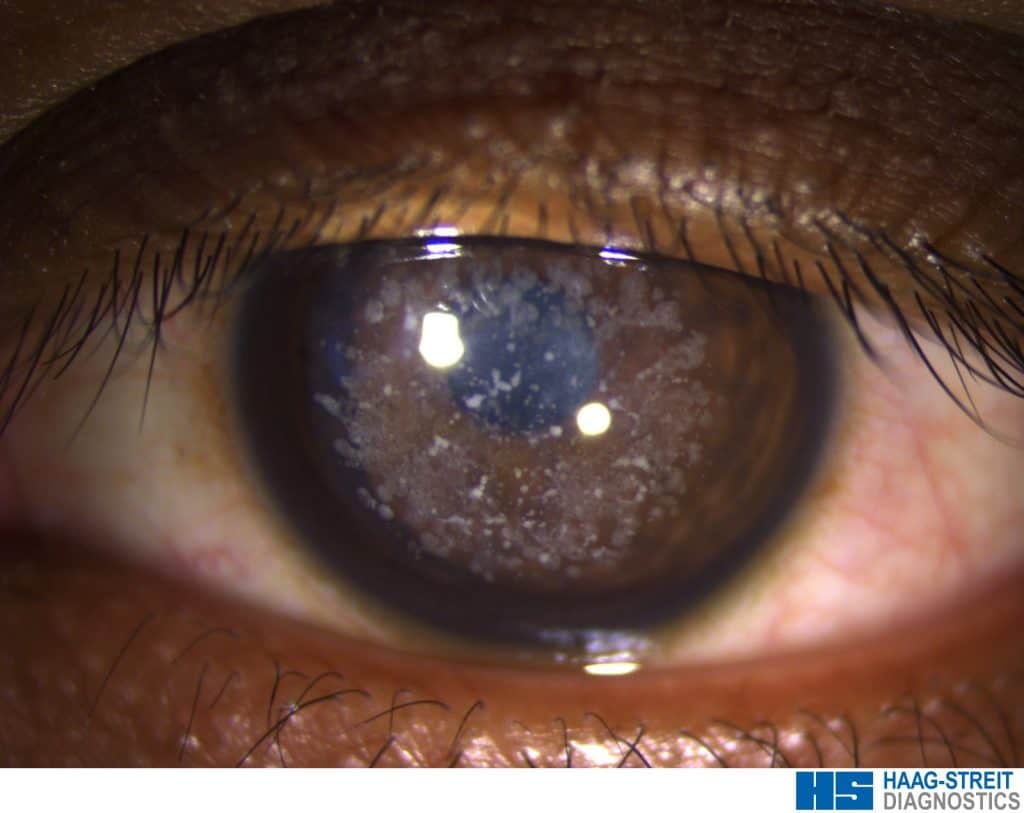
Fig 1: Corneal dystrophy – an inherited disorder of the cornea.
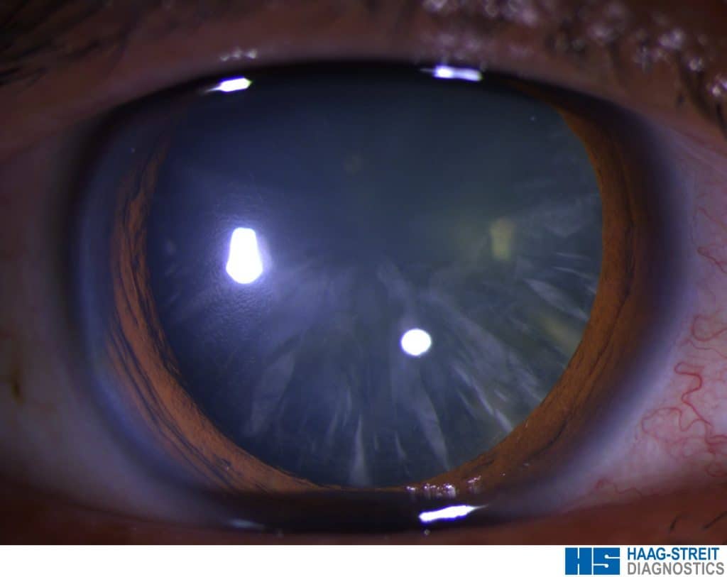
Fig 2: A cortical cataract – cataracts come in different forms, in this type there are radial spokes where the opacity is in the edge of the lens.

Fig 3: The same cataract viewed with retro-illumination – the spokes are easily seen here. When the spokes reach the centre of the lens they can interfere with the passage of light through the lens, resulting in blurred vision.
![]()
Fig 4: Nuclear sclerotic cataract – this is the commonest type of cataract in which the nucleus or centre of the lens turns yellow and eventually brown. This scatters light making the vision blurry and also makes the eye become more myopic (near-sighted).

Fig 5: Punctate epithelial erosions – these are a sign of a dry eye. Fluorescein dye is administered in the form of an eye drop, and when the eye is illuminated with blue light, the dye emits green light. The green dots on the surface of the cornea show dry spots where there is a deficiency of the epithelial cells that form the outermost layer of the cornea. These epithelial erosions usually disappear over time as the dry eye responds to treatment.

Fig 6. Iris neovascularization – this diabetic patient presented with fine new blood vessels growing on his iris. This can result in high eye pressure and a form of glaucoma which can be difficult to treat. The blood vessels completely regressed after eye injections of aflibercept (Eyelea). The photographs allowed his ophthalmologist to monitor his response to treatment.
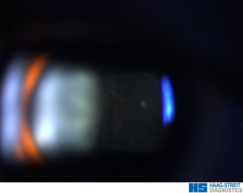
Fig 7. Vitreous cells – this patient has some inflammatory cells (the small white dots in the dark region of the photograph) in the vitreous humor – the transparent gel that fills the eye behind the lens. This is due to inflammation. The photographs are a useful way to measure the response to treatment – the number of cells seen on the photograph should reduce as the treatment takes effect.


