An epiretinal membrane (ERM) is a thin sheet of fibrous tissue that develops on the surface of the macula, the small yellowish area at the centre of the retina. This condition is also known as macular pucker, epimacular membrane, premacular fibrosis, surface wrinkling retinopathy and cellophane maculopathy. Progression of epiretinal membrane in 5 years.

ERM is commonly caused by an ageing or degenerative process called posterior vitreous detachment (PVD). PVD occurs when the jelly-like substance in the eye, known as vitreous humour, shrinks and separates from the retina. This creates a space between the vitreous humour and the retina. As a healing response, certain retinal cells migrate into this space and gradually form an abnormal cellophane-like membrane over the macula. This membrane slowly thickens and contracts over time, resulting in distortion of the retinal blood vessels and wrinkling of the retina. In some cases, the shrinking causes tension and swelling of the macula.
While most cases of ERM are related to ageing, ERM can also occur in certain other eye conditions such as retinal vascular diseases, retinal tear and inflammation of the internal eye structures. Patients with diabetes may develop a condition called diabetic retinopathy which can also cause ERM. In addition, those with eye injuries are also at risk of developing ERM.
An ERM can also occur after retinal operations such as retinal reattachment surgery and laser treatment.
Many people with ERM have no visual symptoms. However, some of them may experience the following symptoms:
- Blurred distant and near vision
- Wavy and distorted vision
- Cloudy or greyish central vision
- Central blind spot
For many patients, the vision tends to remain stable although in some cases, it may gradually worsen over time.
An ERM is usually diagnosed during a retinal examination by an eye or retinal specialist. It can also be diagnosed by taking a colour photograph of the retina using a special camera (retinal photography).
A number of specialised tests such as Serial Colour Retinal Photography, Fundus Autofluorescence Imaging, High-definition optical coherence tomography (OCT), Fundus fluorescein angiography can help your eye or retinal specialist evaluates and manages ERM better.
There is no oral medication or eyedrop available to treat ERM. Depending on the severity of the ERM, it may be observed or treated by surgery.
Observation
In most cases of ERM where there is no or very mild symptom, no treatment is necessary and the condition can be observed. A change in the spectacle prescription may improve the vision slightly.
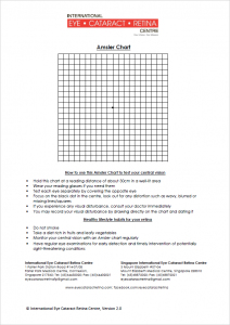
An Amsler grid can be used regularly at home to monitor the central vision for progression of the condition. The patient should be monitored regularly by the eye or retinal specialist with the aid of serial colour retinal photography and high-definition OCT.

A few cases of ERMs have been reported to peel off the macula on their own. However, such spontaneous resolution of ERM is extremely rare.
Surgery
For ERMs causing more severe visual disturbances such as visual distortion or decreased vision, treatment with surgery is recommended. The procedure involves physically removing the membrane in an operation known as vitrectomy and membrane peeling.
In most cases, although visual distortion does improve significantly after surgery, it may not resolve completely and the visual acuity may not return to normal. In general, the visual outcome after surgery tends to be better in those who have mild ERM compared to in those with more severe ERM. Recurrence of ERM may occur in a small proportion of cases.
Potential Risks
Vitrectomy and membrane peeling is a major eye surgery and like any other surgery, there are risks associated with this operation. Some of these risks include the development of cataract, retinal tear, retinal detachment, retinal damage, retinal scarring, severe bleeding, raised eyeball pressure, infection, worsening of vision and total blindness. Major complications occur in less than a few percent of cases.
The retina can become torn during the operation. Should this complication occur, the surgeon will have to repair the retinal tear to prevent the retina from becoming detached. This is usually done using a laser and by injecting a gas bubble into the eye. If a gas bubble is used, the patient will have to keep his head in a certain position after surgery for about 2 to 3 weeks so that the bubble can keep the retina in place while the retinal tear heals. The exact position to place the head after surgery depends on the location of the retinal tear.
Depending on the type of gas used, the gas bubble will disappear on its own in about 1 to 2 months. While the gas bubble remains in the eye, it is unsafe for the patient to fly in an aeroplane as the gas bubble may expand during the flight and cause blindness. If the patient needs to fly soon after the operation, a silicone oil bubble can be used instead of a gas bubble. However, unlike a gas bubble which will gradually disappear on its own, the silicone oil bubble will have to be removed with a minor operation at a later date.
Vitrectomy and membrane peeling can be combined with cataract surgery if the patient also has a cataract. For those who do not have a cataract and have not undergone cataract surgery, a cataract causing impairment of vision will usually form within several months to a few years after the procedure. A cataract surgery will then be required to improve vision. For this reason, some surgeons recommend performing cataract surgery together with the vitrectomy and membrane peeling to save the patient the hassle of another surgery at a later date.
